|
|
|
|
|
A Rare Case of Epidermoid Cyst in Parotid Gland: Case Report and Literature Review
|
|
|
|
Mitsuhiro Aoki1, Hiroshi Okuda2, Natsuko Obara2, Hideki Mori3 Departments of 1Otolaryngology and 3Diagnostic Pathology, Ogaki Tokushukai Hospital; 2Department of Otolaryngology-Head and Neck Surgery, Gifu University Graduate School of Medicine, Ogaki City, Gifu, Japan. |
|
|
|
|
|
Corresponding Author:
|
|
Dr Mitsuhiro Aoki Email: mitsuhiro.aoki@tokushukai.jp |
|
|
|
|
|
|
|
|
Received:
09-NOV-2023 |
Accepted:
18-MAR-2024 |
Published Online:
25-SEP-2024 |
|
|
|
|
|
|
|
Abstract
|
|
|
|
Background: Epidermoid cysts (ECs) are rare, benign lesions that can develop in various areas, including the head and neck, but involvement of the parotid gland is uncommon. Diagnosing them can be difficult due to their cystic nature, often requiring advanced imaging and histopathological analysis. Case Report: A 47-year-old woman presented with a gradually enlarging, mildly painful mass in front of her right ear. Examination revealed a soft, elastic 3 cm mass near the right tragus. Imaging confirmed a well-defined cystic lesion in the superficial lobe of the right parotid gland, and aspiration cytology suggested an epidermoid cyst (EC). The patient underwent a superficial parotidectomy, and the tumor was excised without complications. Histopathology confirmed the diagnosis of EC. Conclusion: Although rare, ECs should be considered in the differential diagnosis of parotid cystic lesions. This case emphasizes the importance of comprehensive imaging and histopathological evaluation in managing cystic parotid masses. |
|
|
|
|
|
Keywords :
|
Epidermoid cysts, Female, Imaging, Parotid Gland.
|
|
|
|
|
|
|
|
|
|
|
|
Introduction
Epidermoid cysts (ECs) in parotid gland are uncommon, and diagnosis may be challenging due to the cystic nature of the lesion, often necessitating imaging and histopathological evaluation [ 1- 3]. Here, we report the case of a 47-year-old woman who presented with a progressively enlarging, soft mass in front of her right ear. Diagnostic investigations, including ultrasonography, computed tomography (CT), and magnetic resonance imaging (MRI), revealed a cystic lesion within the right parotid gland. Surgical excision confirmed the diagnosis of EC. This case highlights the importance of considering ECs in the differential diagnosis of cystic parotid masses and the role of surgery in definitive diagnosis and treatment.
Case Report
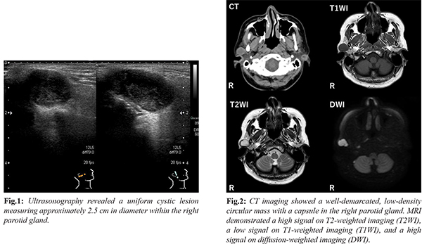
A 47-year-old female presented with a soft mass in front of her right ear, which she had noticed for some time. She sought medical attention as the mass had gradually increased in size and was associated with mild pain. On examination, an elastic, soft mass approximately 3 cm in diameter was palpable in front of the right tragus, with mild tenderness. The patient had no significant medical history, including no prior surgeries in the head and neck region. Ultrasonography revealed a uniform cystic lesion measuring about 2.5 cm in the right parotid gland [Fig.1]. Aspiration cytology showed only viscous fluid. Computed tomography (CT) demonstrated a well-defined, low-density, circular mass with a capsule located in the upper portion of the superficial lobe of the right parotid gland [Fig.2]. Magnetic resonance imaging (MRI) showed a well-circumscribed nodular lesion measuring 29×16×15 mm in the superficial lobe of the right parotid gland. The lesion exhibited uniform signals, with a high signal on T2-weighted imaging (T2WI), a low signal on T1-weighted imaging (T1WI), and a high signal on diffusion-weighted imaging (DWI) [Fig.2]. Based on these findings, we strongly suspected an epidermoid cyst (EC) as the cystic lesion contained viscous fluid. No enlarged lymph nodes were noted around the tumor. The differential diagnosis included lymphoepithelial cysts, non-neoplastic cysts, and complete cystic degeneration of solid tumors such as Warthin's tumor.
After obtaining informed consent, the patient underwent a superficial parotidectomy using a facelift incision, as a neoplastic condition could not be excluded. During surgery, the facial nerve trunk was identified, and its temporal branch was traced. Once it was confirmed that the tumor was separate from the facial nerve, the mass was resected along with part of the parotid gland and surrounding lymphatic tissue. The deep portion of the tumor was located along the anterior superior wall of the external auditory canal and was successfully excised without rupturing the cyst capsule [Fig.3]. Pathological examination revealed two cystic lesions lined with squamous epithelium and containing keratinized material, confirming the diagnosis of ECs [Fig.4]. At the six-month follow-up, the patient showed no signs of local recurrence.
Discussion
Epidermoid cysts (ECs) of the parotid gland are rare and may arise from either congenital or acquired causes. Congenital ECs are remnants of epithelium from embryonic development, while acquired ECs result from epithelial cell implantation due to trauma or surgery [ 4]. Infection and epithelial proliferation following surgical procedures such as myringoplasty or botulinum toxin injection into the masseter muscle have been implicated in acquired cases of ECs in the parotid gland [ 5- 7]. Despite their rarity, few cases of parotid gland ECs have been documented in the English literature [ 1- 10]. In our case, the cyst's location near the external auditory canal suggests a possible congenital origin, potentially linked to a first branchial cleft anomaly, similar to Type I in the Work classification [11]. Type I branchial cleft cysts are commonly found along the external auditory canal, extending into the upper part of the parotid gland. It is likely that in this case, infection or inflammation caused the cyst to enlarge. While most ECs in the head and neck region are benign, there have been reports of malignant transformation into squamous cell carcinoma, especially in intracranial ECs [12,13]. Though no cases of malignant transformation of parotid ECs have been reported [6], the risk factors for malignancy include rapid growth, recurrent lesions, and a cyst size larger than 2 cm [12,14]. Chronic inflammation may play a role in malignant transformation. Diagnosing ECs pre-operatively is challenging due to their cystic nature. Aspiration cytology, though useful, may yield inadequate samples or false negatives, especially in cystic lesions compared to solid tumors [15]. The presence of squamous epithelial cells in aspiration cytology may raise suspicion, but differentiation from malignant tumors or squamous metaplasia is necessary since normal parotid glands lack squamous epithelium. Only about 30% of ECs in the parotid gland are accurately diagnosed pre-operatively [ 8]. Given the potential for malignant transformation in ECs of other regions and the diagnostic challenges, surgical excision remains the preferred treatment for parotid gland ECs [ 12].
Conclusion
Although epidermoid cysts commonly occur in the head and neck skin, their presence within the parotid gland is rare. Our case supports the hypothesis that congenital remnants may enlarge due to infection. While malignant transformation of ECs in other regions has been documented, complete surgical excision is recommended for definitive treatment of parotid gland ECs to prevent potential complications.
Contributors: MA: Concept, design, literature search, manuscript writing; HO: data collection, literature search; NO: data collection, literature search; HM: pathological advice, critical inputs into the manuscript. MA will act as a study guarantor. All authors approved the final version of this manuscript and are responsible for all aspects of this study. Funding: None; Competing interests: None stated.
References - Richardson GS, Clairmont AA, Erickson ER. Cystic lesions of the parotid gland. Plast Reconstr Surg. 1978;61:364-370.
- Devgan M, Devgan BK. Epidermal inclusion cyst in pleomorphic adenoma (mixed parotid tumor). Ear Nose Throat J. 1977;56:75-77.
- Ganesan A, Nandakumar GK. Epidermal cyst of parotid gland: a rarity and a diagnostic dilemma. Case Rep Dent. 2015;2015:856170.
- Hoang VT, Trinh CT, Nguyen CH, Chansomphou V, Chansomphou V, Tran TTT. Overview of epidermoid cyst. Eur J Radiol Open. 2019;6:291-301.
- Thompson AC, Bradley PJ. Iatrogenic epidermoid cyst of the parotid region following ear surgery. J Laryngol Otol. 1991;105:227-228.
- Mahalakshmi S, Reddy S, Ramamurthy TK, Shilpa B. Rare locations of epidermoid cyst: Case reports and review. Ethiop J Health Sci. 2016;26:595-601.
- Ozcan KM, Dere H, Ozcan I, Gun T, Unal T. An epidermal cyst in the parotid gland following ear surgery: a case report. B-ENT. 2006;2:193-195.
- Hegde PN, Prasad HLK, Kumar YS, Sajitha K, Roy PS, Raju M, et al. A rare case of an epidermoid cyst in the parotid gland - which was diagnosed by fine needle aspiration cytology. J Clin Diagn Res. 2013;7:550-552.
- Samia M. Epidermoid cyst of the parotid gland: Report of a rare case with literature review. Otolaryngology (Sunnyvale). 2022;12:3.
- Manz D, Bankfalvi A, Lehnerdt G. Epidermal cyst of the parotid gland. HNO. 2011;59:64-67.
- Work WP. Newer concepts of first branchial cleft defects. Laryngoscope. 1972;82:1581-1593.
- Vellutini EA, de Oliveira MF, Ribeiro AP, Rotta JM. Malignant transformation of intracranial epidermoid cyst. Br J Neurosurg. 2014;28:507-509.
- Solanki SP, Maccormac O, Dow GR, Smith S. Malignant transformation of residual posterior fossa epidermoid cyst to squamous cell carcinoma. Br J Neurosurg. 2017;31:497-498.
- Apollos JR, Ekatah GE, Ng GS, McFadyen AK, Whitelaw SC. Routine histological examination of epidermoid cysts; to send or not to send? Ann Med Surg (Lond). 2017;13:24-28.
- Pantanowitz L, Thompson LDR, Rossi ED. Diagnostic approach to fine needle aspirations of cystic lesions of the salivary gland. Head Neck Pathol. 2018;12:548-561.
|
|
|
|
|
|
|
Search Google Scholar for
|
|
|
Article Statistics |
|
Aoki M, Okuda H, Obara N, Mori HA Rare Case of Epidermoid Cyst in Parotid Gland: Case Report and Literature Review.JCR 2024;14:77-80 |
|
Aoki M, Okuda H, Obara N, Mori HA Rare Case of Epidermoid Cyst in Parotid Gland: Case Report and Literature Review.JCR [serial online] 2024[cited 2025 Mar 13];14:77-80. Available from: http://www.casereports.in/articles/14/3/A-Rare-Case-of-Epidermoid-Cyst-in-Parotid-Gland.html |

|
|
|
|
|