|
|
|
|
|
Giant Vulval Acrochordon: A Common Tumor in a Rare Location
|
|
|
|
Girija BS, Nayanashree V Department of Obstetrics and Gynaecology, Hassan Institute of Medical Sciences, Hassan, Karnataka, India. |
|
|
|
|
|
Corresponding Author:
|
|
Dr Girija BS Email: girijaprasannaobg@gmail.com |
|
|
|
|
|
|
|
|
Received:
09-OCT-2024 |
Accepted:
10-FEB-2025 |
Published Online:
25-MAR-2025 |
|
|
|
|
|
|
|
Abstract
|
|
|
|
Background: Acrochordon, or fibroepithelial polyp, is a benign mesenchymal lesion commonly found on keratinized surfaces, particularly in the axilla and neck. Typically, these lesions measure 3-5 mm. However, vulval occurrence of a giant acrochordon is rare. Case Report: A 26-year-old para 1, living 1 woman presented with a progressively enlarging swelling on the left labia, first noticed three years ago. Initially measuring 2-3 cm, the mass grew to 23×15 cm within a year, leading to discomfort. Examination revealed a multilobulated, pedunculated polypoid mass with ulcerated areas. The lesion was mobile and tender, with no associated vaginal or systemic abnormalities. Surgical excision was performed under spinal anaesthesia, and histopathological examination confirmed the diagnosis of acrochordon. Conclusion: Giant vulval acrochordons can be misdiagnosed as malignant due to their rapid growth and morphological appearance. Histopathology remains the gold standard for diagnosis, and complete surgical excision is curative. |
|
|
|
|
|
Keywords :
|
Excision, Fibroepithelial Neoplasm, Malignant Neoplasms, Pain, Vagina.
|
|
|
|
|
|
|
|
|
|
|
|
Introduction
Acrochordon, commonly known as a skin tag, fibroepithelial polyp, or soft fibroma, is a benign mesenchymal lesion found on keratinized skin surfaces [ 1]. These lesions appear as tan or brown pedunculated papules or nodules ranging from 2-6 mm in size. They commonly occur in obese women of reproductive age, particularly in the neck, axilla, and groin. Genital involvement is rare, with occasional cases reported in the vagina and vulva [ 2]. The hormonal sensitivity of vulvovaginal epithelium may contribute to the enlargement of these lesions. Patients often seek medical attention due to discomfort caused by a hanging mass. This report presents a rare case of a giant vulval acrochordon.
Case Report
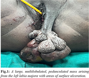
A 26-year-old woman (para 1, living 1) presented with a swelling on the left labia, initially measuring 2-3 cm when first noticed three years ago. Over the past year, it had enlarged to 23×15 cm, causing discomfort. She had no complaints of vaginal discharge, pain, pruritus, abnormal bleeding, or fever. Her menstrual history was normal, and her last childbirth was six years prior. On physical examination, she had a BMI of 31.2 kg/m² (Grade 1 obesity). Systemic examination was unremarkable. Local examination revealed a multilobulated, pedunculated polypoid mass (23×15 cm) arising from the left labia majora, with ulcerated areas on its surface [Fig.1]. The mass was mobile, tender, and varied in consistency. No inguinal lymphadenopathy was noted. Pelvic examination revealed a healthy cervix and vagina, with a normal-sized anteverted uterus and free fornices.

Routine blood investigations were normal. After obtaining informed consent, the mass was excised under spinal anaesthesia using electrocautery. The excised specimen weighed 430 g, with a grey-white gelatinous cut surface containing focal grey-brown and cystic areas. Histopathological examination revealed a polypoidal lesion lined by stratified squamous epithelium with keratinization, fibrocollagenous tissue with an edematous stroma, and scattered spindle cells in a myxoid background with dense neutrophilic infiltration [Fig.2]. Findings confirmed a diagnosis of giant vulval acrochordon. The patient was discharged with a good postoperative outcome but was lost to follow-up.
Discussion
Acrochordons are benign lesions of mesenchymal origin, commonly seen in skin folds like the neck, axilla, and trunk. Perineal involvement is rare. While most lesions measure 2-6 mm, larger ones are uncommon, with some exceeding 2.5 kg in weight [ 3]. Acrochordons have been linked to metabolic disorders, including obesity, hypertension, diabetes mellitus, dyslipidemia, and cardiovascular diseases [ 4]. Their frequent occurrence in reproductive-age and peri-menopausal women, coupled with the presence of estrogen and progesterone receptors in their stromal cells, suggests a hormonal influence. The number and size of acrochordons have been observed to increase during pregnancy and regress postpartum [ 6]. Differential diagnoses for vulval acrochordon include lipoma, fibroma, Bartholin’s cyst, vulval varicosities, hernia, hydrocele of the canal of Nuck, neurofibroma, hemangiomas, angioneurofibroma, hamartoma, lymphadenoma, angiomyofibroblastoma, cellular angiofibroma, sarcomas, angiomyxoma, and dermatofibrosarcoma protuberans [ 2]. Asymptomatic small acrochordons do not require treatment, but larger or symptomatic lesions necessitate removal. Giant acrochordons can cause discomfort while walking and interfere with sexual activity. Surgical excision is the preferred treatment, offering complete resolution with no reported recurrence if excised entirely. However, incomplete excision or multifocality may contribute to recurrence. Rarely, acrochordons may represent an early stage of basal cell carcinoma, emphasizing the need for histopathological confirmation to exclude malignancy [7].
Conclusion
Giant vulval acrochordons are uncommon and can be misdiagnosed as malignant due to their rapid growth and variable morphology. Clinical suspicion, coupled with histopathological examination, is essential for accurate diagnosis and exclusion of neoplastic lesions. Complete surgical excision remains the definitive treatment, ensuring a favourable prognosis with minimal risk of recurrence.
Contributors: GBS: manuscript editing, patient management; NV: manuscript writing, patient management. GBS will act as a study guarantor. Both authors approved the final version of this manuscript and are responsible for all aspects of this study. Funding: None; Competing interests: None stated.
References - Mulik SS, Acharya DB. Large vulval acrochordon in postmenopausal woman - An uncommon presentation: Case Report. J South Asian Feder Obst Gynae. 2023;15(6):749-751.
- Agrawal A, Garg C, Mukherjee S, Mukerjee S, Kakkar KP. Giant acrochordon of vulva: A rare occurrence. Nepal J Dermatology, Venereol Leprol. 2016;13(1):70-72.
- Chaudhury ST. Treatment of unusually large acrochordon by shave excision and electrodesiccation. J Cutan Aesthet Surg. 2008;1(1):21-22.
- Uzuncakmak TK, Akdeniz N, Karadag AS. Cutaneous manifestations of obesity and the metabolic syndrome. Clin Dermatol. 2018;36:81-88.
- Dane C, Dane B, Cetin A, Erginbas M, Tatar Z. Association of psoriasis and vulva fibroepithelial polyp. Am J Clin Dermatol. 2008;9(5):333-335.
- Sharma S, Albertazzi P, Richmond I. Vaginal polyps and hormones - Is there a link? A case series. Maturitas. 2006;53(3):351-355.
- Lortscher DN, Sengelmann RD, Allen SB. Acrochordon-like basal cell carcinomas in patients with basal cell nevus syndrome. Dermatol Online J. 2007;13(2):21.
|
|
|
|
|
|
|
Search Google Scholar for
|
|
|
Article Statistics |
|
Girija BS, Nayanashree VGiant Vulval Acrochordon: A Common Tumor in a Rare Location.JCR 2025;15:24-26 |
|
Girija BS, Nayanashree VGiant Vulval Acrochordon: A Common Tumor in a Rare Location.JCR [serial online] 2025[cited 2025 Apr 24];15:24-26. Available from: http://www.casereports.in/articles/15/1/Giant-Vulval-Acrochordon.html |

|
|
|
|
|