|
|
|
|
|
Dentigerous Cyst Associated with Ectopic Supernumerary Canine in the Maxillary Sinus
|
|
|
claritin and pregnancy claritin and pregnancy
From the Department of ENT Surgery,
University of Maiduguri Teaching Hospital, Maiduguri, Nigeria. |
|
|
|
|
|
Corresponding Author:
|
Dr. YB Ngamdu
Email: ybugam@yahoo.com |
|
|
|
|
|
|
|
|
Received:
24-JUN-2012 |
Accepted:
05-JUL-2012 |
Published Online:
10-JUL-2012 |
|
|
|
|
|
|
|
Abstract
|
|
|
|
Association of ectopic supernumerary tooth in the maxillary sinus with dentigerous cyst is quite uncommon. The cause is unknown in most of the cases but developmental disturbance, pathologic processes or iatrogenic factors have been implicated. Most are symptomless and usually take an insidious course. Those with symptoms will mimic rhinosinusitis. The ectopic tooth can be found incidentally on routine plain radiographs of the paranasal sinuses, skull or panoramic radiographs. We present the case of a patient of large dentigerous cyst associated with an ectopic supernumerary tooth in the right maxillary sinus.
|
|
|
|
|
|
Keywords :
|
Ectopic tooth, Dentigerous cyst, Maxillary sinus.
|
|
|
|
|
|
|
|
|
|
|
|
6go6ckt5b8|3000F7576AC3|Tab_Articles|Fulltext|0xf1ff4464010000003c00000001001400 6go6ckt5b5idvals|133 6go6ckt5b5idcol1|ID 6go6ckt5b5|2000F757Tab_Articles|Fulltext The presence of supernumerary (hyperdontia) or ectopic teeth are not uncommon, and it is estimated to occur in 1% of the general population [1-3]. Ectopic teeth are those that are impacted in unusual positions, or that have been displaced and are at a distance from their normal anatomic location. These ectopic teeth may be permanent, deciduous, or supernumerary [4]. Ectopic eruption of teeth into regions other than the oral cavity has been rarely reported [1,4]. Supernumerary teeth can be classified topographically as mesiodens, paramolar, distomolar and parapremolar. The most common type of supernumerary tooth is mesiodens [5]. The incidence is considerably higher in the maxillary incisor region followed by maxillary third molar and mandibular molar, premolar, canine and lateral incisors [6].
We present our experience of an unusual case that had a large dentigerous cyst associated with ectopic supernumerary canine at roof of right maxillary sinus.
Case Report
A 18 year old male student was referred to the ENT clinic with history of swelling in right cheek following a blunt trauma to right hemiface (hit an electric pole while riding his bicycle) 2 years back.. There was no history of loss of any tooth during trauma. Swelling in the cheek was accompanied by progressive and constant right nasal blockage and headache. Examination revealed a nontender, bony hard mass involving the right check, measuring 8 x 6 cm, completely obstructed right nasal cavity, medial wall of the right maxillary sinus abutting on the septum, fullness of the right hard palate [Figure 1]. Dentition was normal for age.
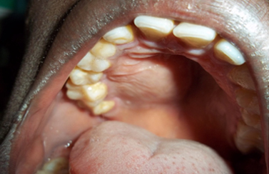 Fig.1: Normal upper jaw dentition, right hard palatal fullness suggestive of dentigerous cyst with intact overlying mucosa.
Water’s view of the paranasal sinuses showed an expanded and complete veiling of the right maxillary sinus with a radiopaque tooth like shadow projecting into the right maxillary sinus at the medial end of the roof [Figure 2].
Fig.1: Normal upper jaw dentition, right hard palatal fullness suggestive of dentigerous cyst with intact overlying mucosa.
Water’s view of the paranasal sinuses showed an expanded and complete veiling of the right maxillary sinus with a radiopaque tooth like shadow projecting into the right maxillary sinus at the medial end of the roof [Figure 2].
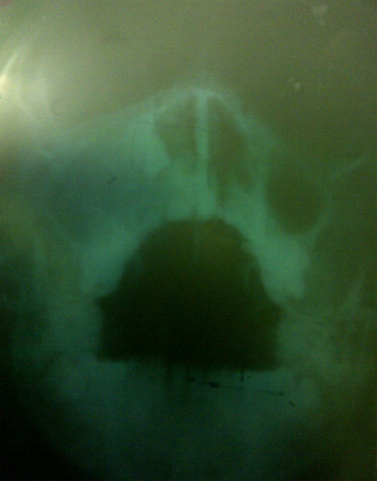 Fig.2: X Ray showing hazy right maxillary sinus with a radio-opaque shadow of tooth at the medial border of the right maxillary sinus.
Caldwell-luc operation under general anaesthesia revealed a cystic mass containing straw coloured fluid and a tooth impacted on the medial end of the roof of the right maxillary sinus. Histology confirmed the wall of the cyst to be dentigerous cyst and the tooth to be canine.
Discussion
Development of tooth progresses through a series of well-defined stages: epithelial thickening, bud, cap and bell [7]. The epithelial thickening expresses a key signalling molecules such as shh that act to increase cell proliferation at the site of the tooth development [8]. The proliferating epithelium invaginates further into the underlying neural crest derived mesenchyme and forms a bud. The instructive information for initiation of a tooth resides in the epithelium, the mesenchyme that starts to condense around the forming bud takes over this instructive role. Thus early oral epithelium and any source of neural crest-derived mesenchyme can form a tooth, and later mesenchyme and a source of non-oral epithelium can also form a tooth [9,10,11]. Abnormal tissue interaction during development may potentially result in ectopic tooth development and eruption [12,13].
Ectopic teeth are those that are impacted in unusual positions or that have been displaced and are at a distance from their normal anatomic location [14]. Ectopic eruption can be associated with developmental disturbance, pathologic process (tumour or cyst) or iatrogenic activity [15,16]. In many cases the aetiology cannot be identified [17]. Ectopic tooth is seen in different regions of the jaw bones such as the mandibular condyle, coronoid process, palate, nasal cavity, places relatively far away from the arch [18,19]. These distally placed teeth are rarely found within the sinus and are discovered on routine clinical or radiographic examination as most cases are asymptomatic [20,21]. Baykal et al reported that of the 14 cases, only one was associated with dentigerous cyst [22]. Ectopic molar and supernumerary teeth have been more commonly reported in the maxillary sinus, but ectopic permanent canines as noted in our case are less frequently encountered [23,24].
Dentigerous cyst is the most common developmental odontogenic cysts, accounting for approximately 25% of all odontogenic cysts of the jaws. They are frequently noted as an incidental finding on radiographs because majority of these cysts are asymptomatic and are most commonly associated with impacted mandibular molar and rarely with permanent maxillary canines [25,26]. Dentigerous cyst usually occurs in the second or third decades of life and rarely seen in childhood [27,28]. Dentigerous cysts are painless in most instances but may cause facial swelling and delayed tooth eruption [27,28]. The dentigerous cyst progress slowly and may exist for several years without being noticed. When the maxillary sinus is invaded, symptoms usually occur late in the process. In other instances, patients become symptomatic and experience the classic features of sinus diseases [29], These may include check swelling [27,30], facial pain [30], headache [29,30], and nasolacrimal obstruction as seen in our patient.
An ectopic tooth in the maxillary sinus is easily diagnosed radiographically, because of their radiopaque image. Water’s view, panoramic radiography and plain skull radiography are simple and inexpensive methods, which can be used in daily practice [31]. The structure of the tooth can be clearly detected on panoramic radiographs. Dentigerous cyst presents as a well-defined radiolucent entity surrounding the crown of an impacted tooth. The border of the cyst is continuous with the cemento-enamel junction of the impacted tooth. This radiographic finding is pathognomonic for dentigerous cyst [26]. Therefore, panoramic radiographs are preferred over CT scan [32]. In the present case, an expanded hazy right maxillary sinus with an ectopic tooth was seen on water’s view. In order to determine the exact location of the ectopic teeth, CT scan would be required. CT scan is necessary and more valuable than plain radiographs not only for definitive diagnosis but also for evaluation of the associated pathology and proper treatment planning. However, in the tropics CT scan is not readily available and affordable, plain radiographs still remain relevant.
The differential diagnosis of a dentigerous cyst includes ameloblastoma, odontogenic fibroma, odontogenic myxoma, reticular cyst, cementomas, pindborg tumour [27]. However, mucoceles, retention cyst and pseudocysts are also considered in the differential diagnosis when a maxillary sinus cyst is visualised involving maxillary sinus expansion[27]. Dentigerous cyst is lined by a layer of nonkeratinized squamous epithelium, with a surrounding wall of a thin connective tissue containing odontogenic epithelial rests [33]. Ameloblastoma or epidermoid carcinomas, occasionally arise from the lining of the dangerous cyst [27,33].
The surgical treatment of an ectopic tooth in the maxillary sinus involves removal via a Caldwell-luc operation [13,16,23]. Although the standard treatment of dentigerous cyst with associated impacted or unerupted tooth is enucleation and extraction of the cyst [28,33]. Marsupialisation is another advisable treatment to preserve the cyst associated tooth and promote its eruption [34], but recurrence and persistence rate is high [35]. Also transnasal extraction of the tooth endoscopically may be attempted if the tooth is small and sited near the ostium of the maxillary sinus [36]. In the present case, a right Caldwell-luc operation was performed to remove the wall of the cyst and the impacted tooth under general anaesthesia. The traditional Caldwell-luc operation provides a direct view into the maxillary sinus; it is associated with more morbidity than trans-nasal endoscopy.
Conclusion
The patient presented here had a rare association of dentigerous cyst with ectopic supernumerary canine in the maxillary sinus. A routine conventional radiograph is the main stay in diagnosing ectopic tooth in the maxillary sinus especially in the tropics where it is readily available and inexpensive. Although when the sinuses are completely opaque, CT scan of the paranasal sinuses is needed to identify location of tooth and other co-existing pathology.
Consent
Written informed consent was obtained from the patient for publication of this case report and any accompanying images.
Competing interests
The authors declare that they have no competing interests
Authors’ contributions
YBN made substantial contribution with regard to the manuscript’s conception and design and was involved in drafting the manuscript. AMK and BMS have made substantial contributions in drafting the manuscript; HIG and AI were involved in revising the manuscript critically. The corresponding author will give final approval of the version to be published.
References
Fig.2: X Ray showing hazy right maxillary sinus with a radio-opaque shadow of tooth at the medial border of the right maxillary sinus.
Caldwell-luc operation under general anaesthesia revealed a cystic mass containing straw coloured fluid and a tooth impacted on the medial end of the roof of the right maxillary sinus. Histology confirmed the wall of the cyst to be dentigerous cyst and the tooth to be canine.
Discussion
Development of tooth progresses through a series of well-defined stages: epithelial thickening, bud, cap and bell [7]. The epithelial thickening expresses a key signalling molecules such as shh that act to increase cell proliferation at the site of the tooth development [8]. The proliferating epithelium invaginates further into the underlying neural crest derived mesenchyme and forms a bud. The instructive information for initiation of a tooth resides in the epithelium, the mesenchyme that starts to condense around the forming bud takes over this instructive role. Thus early oral epithelium and any source of neural crest-derived mesenchyme can form a tooth, and later mesenchyme and a source of non-oral epithelium can also form a tooth [9,10,11]. Abnormal tissue interaction during development may potentially result in ectopic tooth development and eruption [12,13].
Ectopic teeth are those that are impacted in unusual positions or that have been displaced and are at a distance from their normal anatomic location [14]. Ectopic eruption can be associated with developmental disturbance, pathologic process (tumour or cyst) or iatrogenic activity [15,16]. In many cases the aetiology cannot be identified [17]. Ectopic tooth is seen in different regions of the jaw bones such as the mandibular condyle, coronoid process, palate, nasal cavity, places relatively far away from the arch [18,19]. These distally placed teeth are rarely found within the sinus and are discovered on routine clinical or radiographic examination as most cases are asymptomatic [20,21]. Baykal et al reported that of the 14 cases, only one was associated with dentigerous cyst [22]. Ectopic molar and supernumerary teeth have been more commonly reported in the maxillary sinus, but ectopic permanent canines as noted in our case are less frequently encountered [23,24].
Dentigerous cyst is the most common developmental odontogenic cysts, accounting for approximately 25% of all odontogenic cysts of the jaws. They are frequently noted as an incidental finding on radiographs because majority of these cysts are asymptomatic and are most commonly associated with impacted mandibular molar and rarely with permanent maxillary canines [25,26]. Dentigerous cyst usually occurs in the second or third decades of life and rarely seen in childhood [27,28]. Dentigerous cysts are painless in most instances but may cause facial swelling and delayed tooth eruption [27,28]. The dentigerous cyst progress slowly and may exist for several years without being noticed. When the maxillary sinus is invaded, symptoms usually occur late in the process. In other instances, patients become symptomatic and experience the classic features of sinus diseases [29], These may include check swelling [27,30], facial pain [30], headache [29,30], and nasolacrimal obstruction as seen in our patient.
An ectopic tooth in the maxillary sinus is easily diagnosed radiographically, because of their radiopaque image. Water’s view, panoramic radiography and plain skull radiography are simple and inexpensive methods, which can be used in daily practice [31]. The structure of the tooth can be clearly detected on panoramic radiographs. Dentigerous cyst presents as a well-defined radiolucent entity surrounding the crown of an impacted tooth. The border of the cyst is continuous with the cemento-enamel junction of the impacted tooth. This radiographic finding is pathognomonic for dentigerous cyst [26]. Therefore, panoramic radiographs are preferred over CT scan [32]. In the present case, an expanded hazy right maxillary sinus with an ectopic tooth was seen on water’s view. In order to determine the exact location of the ectopic teeth, CT scan would be required. CT scan is necessary and more valuable than plain radiographs not only for definitive diagnosis but also for evaluation of the associated pathology and proper treatment planning. However, in the tropics CT scan is not readily available and affordable, plain radiographs still remain relevant.
The differential diagnosis of a dentigerous cyst includes ameloblastoma, odontogenic fibroma, odontogenic myxoma, reticular cyst, cementomas, pindborg tumour [27]. However, mucoceles, retention cyst and pseudocysts are also considered in the differential diagnosis when a maxillary sinus cyst is visualised involving maxillary sinus expansion[27]. Dentigerous cyst is lined by a layer of nonkeratinized squamous epithelium, with a surrounding wall of a thin connective tissue containing odontogenic epithelial rests [33]. Ameloblastoma or epidermoid carcinomas, occasionally arise from the lining of the dangerous cyst [27,33].
The surgical treatment of an ectopic tooth in the maxillary sinus involves removal via a Caldwell-luc operation [13,16,23]. Although the standard treatment of dentigerous cyst with associated impacted or unerupted tooth is enucleation and extraction of the cyst [28,33]. Marsupialisation is another advisable treatment to preserve the cyst associated tooth and promote its eruption [34], but recurrence and persistence rate is high [35]. Also transnasal extraction of the tooth endoscopically may be attempted if the tooth is small and sited near the ostium of the maxillary sinus [36]. In the present case, a right Caldwell-luc operation was performed to remove the wall of the cyst and the impacted tooth under general anaesthesia. The traditional Caldwell-luc operation provides a direct view into the maxillary sinus; it is associated with more morbidity than trans-nasal endoscopy.
Conclusion
The patient presented here had a rare association of dentigerous cyst with ectopic supernumerary canine in the maxillary sinus. A routine conventional radiograph is the main stay in diagnosing ectopic tooth in the maxillary sinus especially in the tropics where it is readily available and inexpensive. Although when the sinuses are completely opaque, CT scan of the paranasal sinuses is needed to identify location of tooth and other co-existing pathology.
Consent
Written informed consent was obtained from the patient for publication of this case report and any accompanying images.
Competing interests
The authors declare that they have no competing interests
Authors’ contributions
YBN made substantial contribution with regard to the manuscript’s conception and design and was involved in drafting the manuscript. AMK and BMS have made substantial contributions in drafting the manuscript; HIG and AI were involved in revising the manuscript critically. The corresponding author will give final approval of the version to be published.
References
- Moreano EH, Zich DK, Goree JC, Graham SM. Nasal tooth. Am J Otolaryngol. 1998;19:124–126.
- Thor AL. Delayed removal of a fully intruded primary incisor through the nasal cavity: A case report. Dent Traumatol 2002;18:227–230.
- Wang LF, Tai CF, Lee KW, Ho KY, Kuo WR. Delayed removal of a fully intruded tooth in the nasal cavity after facial trauma: A case report. Otolaryngol Head Neck Surg. 2004;131:330–331.
- Carver DD, Peterson S, Owens T. Intranasal teeth: A case report. Oral Surg Oral Med Oral Pathol. 1990;70:804–805.
- Alberti G, Mondani PM, Parodi V. Eruption of supernumerary permanent teeth in a sample of urban primary school population in Genoa, Italy. Eur J Paediatr Dent 2006 ;7:89-92.
- Grimanis GA, Kyriakides AT, Spyropoulos ND. A survey on supernumerary molars. Quintessence Int. 1991;22:989–995.
- Javier C, Abigail ST. Current knowledge of tooth development: Paterning and mineralization of the murine dentition .J Anat. 2009;214:502-515.
- Hardcastle Z, Mo R, Lui CC, Sharpe PT. The Shh signalling pathway in tooth development: defects in Gli2 and Gli3 mutants. Development 1998;125:2803-2811.
- Mina M, Kollar EJ. The induction of odontogenesis in non dental mesenchyme combined with early murine mandibulat arch epithelium. Arch Oral Biol. 1987;32:123-127.
- Lumsden AG. Spatial organization of the epithelium and the role of nueral crest cells in the initiation of mammalian tooth germ. Development 1988;103:155-169.
- Ruch JV, Lesot H, Karcher-Djuricic V, Meyer MJ.Extracellular matrix-mediated interaction during odontogenesis. Prog Clin Biol Res. 1984;151:103-114.
- Srinivasa PT, Sujatha G, Niazi TM, Rajesh P. Dentigerous cyst associated with ectopic third molar in the maxillary sinus: a rare entity. Indian J Dent Res. 2007;18:141-143.
- Goh YH. Ectopic eruption of maxillary molar tooth-an unusual cause of recurrent sinusitis. Singapore Med J. 2001;42:80-81.
- Nagarajappa D, Manjanatha BS. Tooth in oropharynx. J Oral Maxillo Fac Path. 2011;15:346-347.
- Büyukkurt MC, Tozoglu S, Aras MH, Yolcu U. Ectopic eruption of a maxillary third molar tooth in the maxillary sinus: A case report. J Contemp Dent Pract. 2005;6:104–110.
- Bodner L, Tovi F, Bar-Ziv J. Teeth in the maxillary sinus–imaging and management. J Laryngol Otol. 1997;111:820–824.
- Chye CH, Singh B. Rapid cystic development in relation with an impacted lower third molar: A case report. Ann Acad Med Singapore 2005;34:130–133.
- Laskaris G. Color atlas of oral diseases in children and adolescents. Thieme Stuttgart 2000;12-13.
- El-Sayed Y. Sinonasal teeth. J Otolaryngol 1995;24:180-183.
- Elonga S, Palaniappa SP. Ectopic tooth in the roof of maxillary sinus. Ear Nose Throat J. 1991;70:3365-3366.
- Yeung kh, Lee KH. Intranasal tooth in a patient with cleft lip and alveolus. Cleft Palate Creniofac J 1996;33:157-159.
- Baykul T, Dogru H, Yasan H, Aksoy MC. Clinical impact of ectopic teeth in maxillary sinus. Auris Nasus Larynx 2006;33:277-281.
- Atlas E, Karasen RM, Yilmaz AB, Aktan B, Kocer I, Erman Z. A case of a large dentigerous cyst containing a caninein the maxillary antrum leading to epiphora. J Laryngol Otol. 1997;111:641-643.
- Edwards JL, Ferguson JW. Ectopic maxillary canine. N Z Dent J. 1976;72:33-34.
- Daley TD, wysocki GP, Pringle GA. Relative incidence of odontogenic tumor and oral and jaw cysts in a Canadian population. Oral Surg Oral Med Oral Pathol. 1994;77:276-280.
- White SC, Pharoah MJ. Cysts and cystlike lesions of the jaws. White SC, Pharoah MJ, St Louis MO In: Oral Radiology principle and interpretation, 6th ed. Mosby Elsevier; 2009;346-350.
- Ustuner E, Fitoz S, Atasoy C, Erden I. Bilateral maxillary dentigerous cyst: A case report. Oral Surg Oral Med Oral Pathol Oral Radiol Endod. 2003;95:632-635.
- Smith JL 2nd, Kellman RM. Dentigerous cysts presenting as head and neck infections. Otolaryngol Head Neck Surg. 2005;133;715-717.
- Freeland ES, Henneman PL. An unusual cause of headache: A dentigerous cyst in the maxillary sinus. Ann Emerg Med. 1987;16:1174-1176.
- Miconzkadioglu SD, Erkan AN. Endoscopic removal of maxillary dentigerous cyst. B-ENT. 2007;3:213-216.
- Saiki T, Yumoto E. Quantification of x-ray opacity of the maxillary sinus in water`s view. Auris Nasus Larynx 1997;24:289-297.
- Saadattin D, Binali C, Mustafa G. A dentigerous cyst containing an ectopic canine tooth below the floor of the maxillary sinus: A case report.J Oral Science 2007;49:249-252.
- Tournas AS, Tewfik MA, Chauvin PJ, Manoukian JJ. Multiple unilateral maxillary dentigerous cysts in a nonsyndromic patient: A case report and review of the literature. J Oral Maxillofac Surg. 1998;56:237-279.
- Hyomoto M, Kawakami M, Inoue M, Kirita T. Clinical conditions for eruption of maxillary canines and mandibular premolars associated with dentigerous cysts. Am J Orthod Dentofacial Orthop. 2003;124:515-520.
- Ertas U, Yavuz MS. Interesting eruption of 4 teeth associated with large dentigerous cyst in mandible by only marsipialisation. J Oral Maxillofac Surg. 2003;61:728-730.
- Goh YH. Ectopic eruption of maxillary molar tooth-an unusual cause of recurrent sinusitis. Singapore Med J 2001;42:80-81.
|
|
|
|
|
|
|
Search Google Scholar for
|
|
|
Article Statistics |
|
Ngamdu YB, Kodiya AM, Sandabe MB, Garandawa HI, Isa ADentigerous Cyst Associated with Ectopic Supernumerary Canine in the Maxillary Sinus.JCR 2012;2:27-30 |
|
Ngamdu YB, Kodiya AM, Sandabe MB, Garandawa HI, Isa ADentigerous Cyst Associated with Ectopic Supernumerary Canine in the Maxillary Sinus.JCR [serial online] 2012[cited 2024 Apr 16];2:27-30. Available from: http://www.casereports.in/articles/2/2/dentigerous-cyst-associated.html |

|
|
|
|
|